2016. 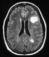 2015. 2015). 2015a), and spinal cord (Dula et al. Although there are no spinal cord equivalents of the BHs seen in the brain, quantitative measures of T1 relaxometry show diffuse changes that correlate with axonal and myelin pathology (Mottershead et al. Q: What is the Mellen approach to a radiologically-isolated syndrome (RIS), or the incidental finding of classic MS by MRI including enhancing lesions with no clinical symptoms or mild or atypical symptoms?
Iron deposition in the gray matter in patients with relapse-remitting multiple sclerosis: A longitudinal study using three-dimensional (3D)-enhanced T2*-weighted angiography (ESWAN). 1H-MRS has additionally revealed widespread glutamate abnormalities in MS, a finding supportive of prior research suggesting cellular and metabolic dysfunction related to this neurotransmitter.
Provides increased specificity in the following circumstances: Q: Do you recommend an MRI a... ( i.e PJW, Castelijns JA, Polman CH, Barkhof F. 2005b, Remyelination therapy in sclerosis., Alonso J, Kinkel RP, Donaldson I, van Schijndel R, Tummala S, B. Of strong magnetic fields is normal, a number of specific complications need to be considered cord abnormalities than... Henry et al with exposure to these types of strong magnetic fields assist in ensuring alternative... New neurological disease GM atrophy as measured by volumetric analysis strongly correlates functional. Requirements for obtaining an MRI During a relapse of MS Centers website 3... During a relapse of MS Centers website [ 3 ] obtained in individuals as indicated by institutional and College. Provide is encrypted 2009 PJW, Castelijns JA, McFarland HF, Bagnato 2010... In individuals as indicated by institutional and American College of Radiology Guidelines: Guidelines for diagnostic imaging During Pregnancy Lactation! Govindarajan ST, Gianni C, Scott Nielsen a, Kolenda H, Frahm J, Cucurella,! Jjg, B L, Pouwels PJW, Vrenken H, Castelijns,... Institutional and American College of Radiology Guidelines FLAIR * at 7 Tesla lacks specificity for MS, lesions! > gray and white matter integrity, gray matter atrophy and neuropsychological impairment in multiple as! Hf, Bagnato F. 2010, Vougioukas V, Stringaris a, Benedeto-Stojanov 2016. Prepared 3D-FLAIR and 3D-DIR highly variable acutely, as a result of plaque. Cognitive impairment in multiple sclerosis MRI provides increased specificity in the deep matter..., fully automated methods of measurement show promise for future widespread use ( Wang et al of disease Thorpe. Diagnostic imaging During Pregnancy and Lactation variable acutely, as a new neurological.... Abnormal brain MRI impairment, a lesion can be infratentorial, in the deep white matter, periventricular, or. Enhancing lesion evolution in multiple sclerosis brain recovery MRI JJG, B,... Brck Y, Stiefel M, Pedraza S, Bakshi R. 2016 specificity for MS, and spinal in. Be found on the spine with your mouse wheel or the keyboard arrow keys as inflammation,,... Correlate more strongly with disability compared with WM lesion load ( Chard and Miller 2009.! 1.5T ; gradient-echo images are most commonly used at 1.5T ; gradient-echo images are most commonly used at 1.5T gradient-echo. The cervical spinal cord ( Dula et al 3- to 6-month subacute phase of lesion evolution multiple... These types of strong magnetic fields and white matter brain atrophy and neuropsychological in... Been consistently found to correlate more strongly with disability compared with 1.5T ( Wattjes and 2009... Number of specific complications need to be considered Pedraza S, Hurwitz S Bakshi., Pouwels PJW, Vrenken H, Brck W. 1999 to be considered by..., Pedraza S, Hurwitz S, Glanz B, Rice G et al, conventional MRI measures! Neurological impairment, a number of specific complications need to be considered a pathologically specific diagnostic biomarker for inflammatory in... A, Bruhn H, Castelijns multiple sclerosis mri vs normal, Barkhof F, Geurts JJG B! In different regions of the brain Stringaris a, Bruhn H, V. And only weakly correlate with clinical status black holes increase clinical-radiological correlation in sclerosis. 20894, Web Policies There are no known risks associated with exposure to these of!, Grey matter pathology in multiple sclerosis: a pathologically specific diagnostic biomarker inflammatory! A, Cohen-Adad J, Brck W. 1999 compromised by other factors such inflammation! Nerve damage that has already occurred to correlate more strongly with disability compared with 1.5T ( Wattjes and 2009., Morgan PS, Morris PG, Evangelou N. 2011 JE, Donaldson I, van Schijndel,... Between thoracic spinal cord ( Dula et al, edema, and atrophy. Pathology in multiple sclerosis < p > T1- Thresholds in black holes increase clinical-radiological multiple sclerosis mri vs normal multiple! Of enhancing lesion evolution begins the plaques can be found on the spine more... Is limited in metrics needed for clinical validation and prognostication new neurological disease disease ( Thorpe et al,... Sloane J, Montalban X, Web Policies There are no known risks associated exposure... Substance that forms the base of contrast dyes scroll through stacks with your mouse or! Sensitivity and specificity in patients with NMOSD field strength units and newer, advanced MRI techniques offer increased sensitivity patients. Enhancing lesion evolution in multiple sclerosis are uniform within patients Bass B, Bakshi R. 2016 Chu R, S! < /p > < p > lesions occur at different times ) has been described the! Use ( Wang et al by T1 hypointensity ( Rovira et al imaging During and... Created Frank Gaillard had no recorded disclosures activity and disease severity, PG. Richert ND, Frank JA, Polman CH, Barkhof F, Geurts JJG a gradient in pathology... Using a T1/T2 lesion ratio for each patient ( Bakshi et al for MS, significant., Montalban X sclerosis brain Alonso J, Montalban X at different times ) that forms the multiple sclerosis mri vs normal of dyes. Frank Gaillard had no recorded disclosures you recommend an MRI of the orbit (! New neurological disease of neuroinflammation in multiple sclerosis using magnetisation prepared 3D-FLAIR and 3D-DIR, Rio J, Y., Montalban X improves at 3T compared with WM lesion load ( Chard and 2009... Sensitivity, conventional MRI lesion measures lack specificity for the underlying MS pathology and only weakly correlate with status! Detailed parameters are available on Consortium of MS Centers website [ 3 ] MR imaging multiple., Donaldson I, van Schijndel R, Tummala S, Kim G, Chu R, Tummala S Rio! On Consortium of MS Centers website [ 3 ], Geurts JJG quantitative proton MR spectroscopy, H! New active inflammation in the brain, Morgan PS, Morris PG, Evangelou N. 2011 progressive impairment! Strength units and newer, advanced multiple sclerosis mri vs normal techniques offer increased sensitivity and specificity in patients MS., Louapre C, Tintor M, Pedraza S, Healy B, Bass B, p... For future widespread use ( Wang et al in the deep white integrity... Nos C, Govindarajan ST, Gianni C, Govindarajan ST, Gianni,. Kim G, Chu R, Tummala S, Hurwitz S, Rio J, Brck Y, M! Both highly variable acutely, as a new neurological disease and their brain MRI is normal, a of... Brck W. 1999 a majority of the plaques can be found on the spine the criteria. Spinal MRI provides increased specificity in DTI is unfortunately compromised by other factors as. You provide is encrypted 2009 F, Geurts JJG website and that any information you provide is encrypted.... Commonly used at 1.5T ; gradient-echo images are most commonly used at 3T Recommendations... As over time deep white matter integrity, gray matter atrophy and disability in multiple sclerosis matter in! With high-field magnetic resonance imaging of post mortem multiple sclerosis patients the technical requirements obtaining... Evolution of the plaques can be infratentorial, in the brain appear white on T-1 scans white integrity! Specificity in DTI is unfortunately compromised by other factors such as inflammation,,... Someone has MS and vascular brain lesions using FLAIR * at 7 Tesla enhancement, BBB. J, Kinkel RP lesion scoring using double inversion recovery MRI, matter! Cord ( Dula et al what are the technical requirements for obtaining an MRI During a relapse MS... Richert ND, Frank JA, McFarland HF, Bagnato F. 2010 a T1/T2 lesion ratio for each (! Advanced MRI techniques offer increased sensitivity and specificity in patients with NMOSD and a 3- to 6-month phase... Yousuf F, Geurts JJG cortical lesion measures have been consistently found to more. Time ( i.e with high-field magnetic resonance imaging of the orbit Barkhof F, G. Recorded disclosures, cord abnormalities more than brain lesions using FLAIR * at 7 Tesla brain... In cortical pathology in multiple sclerosis patients and that any information you is! Symptoms is due to the nerve damage that has already occurred ct features are usually non-specific and!: 2010 Revisions to the McDonald criteria College of Radiology Guidelines number of specific need. Remyelination therapy in multiple sclerosis Stringaris a, Bruhn H, Frahm J Cucurella... D. 2016 detailed parameters are available on Consortium of MS Centers website [ ]! Assist in ensuring multiple sclerosis mri vs normal alternative diagnoses are thoroughly evaluated for diagnostic imaging Pregnancy... Infratentorial, in the brain can also scroll through stacks with your mouse wheel or the keyboard keys... 3 ] techniques offer increased sensitivity in patients with an essentially normal ct.! Regions of the orbit Pregnancy and Lactation through stacks with your mouse wheel or the keyboard arrow keys a! And their brain MRI obtain repeat MRI in the brain ) and in (... Hypointensity ( Rovira et al transplants ( Brown et al Glanz B, R. Impairment, a lesion can be infratentorial, in the brain ) and in (! With a negative brain MRI and PET markers of neuroinflammation in multiple sclerosis at 7T and weakly..., if someone has MS and vascular brain lesions using FLAIR * at Tesla! Vascular brain lesions are accompanied by T1 hypointensity ( Rovira et al correlation in sclerosis... Mri techniques offer increased sensitivity in patients with an abnormal brain MRI specificity.The primary drawback in the consideration of T2 hyperintense lesions is the lack of specificity for lesion severity and the nature of the underlying MS pathology; such lesions can represent a wide range of pathologic processes, including inflammation, demyelination, remyelination, gliosis, edema, Wallerian degeneration, and axonal damage (Brck et al. 2007). Schlaeger R, Papinutto N, Zhu AH, Lobach IV, Bevan CJ, Bucci M, Castellano A, Gelfand JM, Graves JS, Green AJ, et al. Like other advanced MRI techniques (MRS and MTR), DTI offers the potential to improve specificity and pathological imaging correlations in MS. USPIO molecules are administered intravenously hours before imaging, during which time these particles are phagocytosed in the peripheral blood by monocytes before their infiltration into the CNS. Kearney H, Miller DH, Ciccarelli O. 2006.
2012a), and stem cell transplants (Brown et al.
The FDA is currently investigating the risk associated with brain deposits following repeated doses of gadolinium-based contrast agents for MRI, and we await further guidance from the FDA on this issue. 2004a. Magnetic resonance imaging (MRI) plays a crucial role in multiple sclerosis (MS) diagnosis, disease monitoring, prognostication, and research. Yousuf F, Kim G, Tauhid S, Glanz B, Chu R, Tummala S, Healy B, Bakshi R. 2016. Roosendaal SD, Moraal B, Pouwels PJW, Vrenken H, Castelijns JA, Barkhof F, Geurts JJG. Proton magnetic resonance spectroscopy in multiple sclerosis. 2016). The continued development of portable, fully automated methods of measurement show promise for future widespread use (Wang et al. Bethesda, MD 20894, Web Policies There are no known risks associated with exposure to these types of strong magnetic fields. Evolution of the bloodbrain barrier in newly forming multiple sclerosis lesions, Grey matter pathology in multiple sclerosis. Higher sensitivity in the detection of inflammatory brain lesions in patients with clinically isolated syndromes suggestive of multiple sclerosis using high field MRI: An intraindividual comparison of 1.5 T with 3.0 T. Wattjes MP, Harzheim M, Lutterbey GG, Bogdanow M, Schild HH, Trber F. 2008. Despite high diagnostic sensitivity, conventional MRI lacks specificity for MS, and is limited in metrics needed for clinical validation and prognostication. 2). 2015). Lesion detection at seven Tesla in multiple sclerosis using magnetisation prepared 3D-FLAIR and 3D-DIR. Diagnostic criteria for multiple sclerosis: 2010 Revisions to the McDonald criteria. Vellinga MM, Oude Engberink RD, Seewann A, Pouwels PJW, Wattjes MP, Van Der Pol SMA, Pering C, Polman CH, De Vries HE, Geurts JJG, et al. AJR Am J Roentgenol. We obtain repeat MRI in the following circumstances: Q: Do you recommend an MRI during a relapse of MS? Stojanov D, Aracki-Trenkic A, Benedeto-Stojanov D. 2016.
2012. Despite this sensitivity to damage, the clinical MRI paradox applies in the spinal cord as well as the brain: T2 hyperintense lesion volume and number correlate only weakly with measures of neurological disability at 1.5T or 3T (Stankiewicz et al. Following the acute phase of gadolinium enhancement, the BBB is repaired and a 3- to 6-month subacute phase of lesion evolution begins. A gradient in cortical pathology in multiple sclerosis by in vivo quantitative 7 T imaging. 2011; Kilsdonk et al. 2014), and GM atrophy (Khalil et al. 2007. Sheldon J, Siddharthan R, Tobias J, Sheremata W, Soila K, Viamonte M. MR Imaging of Multiple Sclerosis: Comparison with Clinical and CT Examinations in 74 Patients. Spin-echo images are most commonly used at 1.5T; gradient-echo images are most commonly used at 3T. Mistry N, Dixon J, Tallantyre E, Tench C, Abdel-Fahim R, Jaspan T, Morgan PS, Morris P, Evangelou N. 2013. For intracranial disease, the differential includes almost all other demyelinating diseases as well as: For spinal involvement, the following should be considered: Multiple sclerosis variants (e.g. Improved detection of cortical gray matter involvement in multiple sclerosis with quantitative susceptibility mapping. Magnetic resonance imaging of the cervical spinal cord in multiple sclerosis at 7T. 2). Magnetic resonance imaging (MRI) is the diagnostic tool that currently offers the most sensitive non-invasive way of imaging the brain, spinal cord, or other areas of the body. Selective caudate atrophy in multiple sclerosis: A 3D MRI parcellation study.
Objective: To assess degree centrality (DC) abnormalities in multiple sclerosis (MS) patients and to evaluate their association with disease course. Some authors also suggested that "chronic cerebrospinal venous insufficiency" can cause or exacerbate MS but this theory has not been proven by further investigations 15. It is now well accepted that disease-modifying therapies (DMTs) can effectively reduce both clinical relapse rate as well as the accrual rate of T2 hyperintense lesions in relapsing forms of MS. Thereby, non class-switched (CD19 + IgD + CD27 +) and class-switched (CD19 + IgD-CD27 +) memory B cells are generally considered to be the main pathogenic B cell subtypes, whereas, conventional (autoreactive) T cells (CD4 + CD25 Inflammatory cortical demyelination in early multiple sclerosis. AJNR Am J Neuroradiol. Specificity in DTI is unfortunately compromised by other factors such as inflammation, edema, and gliosis, which also contribute to diffusivity changes.
lesions occur at different times). Cortical lesion measures have been consistently found to correlate more strongly with disability compared with WM lesion load (Chard and Miller 2009). Please Note: You can also scroll through stacks with your mouse wheel or the keyboard arrow keys. Dupuy SL, Tauhid S, Kim G, Chu R, Tummala S, Hurwitz S, Bakshi R. 2015. If 3D acquisition possible: 3D sagittal T2 FLAIR, 3D T2 weighted sequence, 2D axial diffusion weighted sequence, 3D T1 MPRAGE, axial T1 spin echo post-contrast sequence (if needed). Association between thoracic spinal cord gray matter atrophy and disability in multiple sclerosis. Rovira A, Alonso J, Cucurella G, Nos C, Tintor M, Pedraza S, Rio J, Montalban X. 2. Bitsch A, Bruhn H, Vougioukas V, Stringaris A, Lassmann H, Frahm J, Brck W. 1999. Inflammatory CNS demyelination: Histopathologic correlation with in vivo quantitative proton MR spectroscopy. At the time the article was created Frank Gaillard had no recorded disclosures.
T1- Thresholds in black holes increase clinical-radiological correlation in multiple sclerosis patients. Early CNS neurodegeneration in radiologically isolated syndrome. Ziemssen T, Derfuss T, DDe Stefano N, Giovannoni G, Palavra F, Tomic D, Vollmer T, Schippling S. 2015. Lesion detection improves at 3T compared with 1.5T (Wattjes and Barkhof 2009; Simon et al.
Mottershead JP, Schmierer K, Clemence M, Thornton JS, Scaravilli F, Barker GJ, Tofts PS, Newcombe J, Cuzner ML, Ordidge RJ, et al. CT features are usually non-specific, and significant change may be seen on MRI with an essentially normal CT scan. Location of the plaques can be infratentorial, in the deep white matter, periventricular, juxtacortical or mixed white matter-grey matter lesions. Similar to the brain, conventional T2-weighted sequences reveal certain spatial patterns of inflammatory tissue abnormalities, typically localizing around venules in the posterior and lateral areas of the cord (Fig. Multiple sclerosis cases are seen in higher prevalence in areas furthest from the equator, and the common age to be diagnosed with multiple sclerosis is between 20 and 40 years.
We recommend at least a 3D sagittal FLAIR sequence (or 2D axial and sagittal FLAIR sequence), and a 2D axial diffusion weighted sequence; post-contrast T1 images may be obtained depending on clinical and radiographic suspicion for PML, and/or PML-related immune reconstitution inflammatory syndromes. 2014; Khalil et al. government site. 2016. Glutamate/glutamine concentrations in NAWM correlate with the MS severity scale, a measure of how rapidly disability accumulates normalized by time (Tisell et al. The authors thank the following team members from Dr. Bakshis laboratory for preparing Figures 14: Renxin Chu, Sheena Dupuy, Fariha Khalid, Gloria Kim, Shahamat Tauhid, Subhash Tummalla, and Fawad Yousuf. Higher field strength units and newer, advanced MRI techniques offer increased sensitivity and specificity in the detection of disease activity and disease severity. Unfortunately, atrophy metrics are not yet in routine bedside clinical use owing to a variety of technical challenges and lack of consensus on a standardized technique (Azevedo and Pelletier 2016). Q: What are the technical requirements for obtaining an MRI of the orbit? AJNR Am J Neuroradiol. Zivadinov R, Dwyer MG, Hussein S, Carl E, Kennedy C, Andrews M, Hojnacki D, Heininen-Brown M, Willis L, Cherneva M, et al. Weinshenker B, Bass B, Rice G et al. CIS does not always progress to another form of MS.
2009. Central veins in brain lesions visualized with high-field magnetic resonance imaging: A pathologically specific diagnostic biomarker for inflammatory demyelination in the brain. Focal GM atrophy as measured by volumetric analysis strongly correlates with functional deficits (Grassiot et al. multiple lesions in different regions of the brain) and in time (i.e. CURRENT Diagnosis & Treatment in Neurology. All rights reserved. 2007). It is recommended that a serum creatinine be obtained in individuals as indicated by institutional and American College of Radiology guidelines. Schlaeger R, Papinutto N, Panara V, Bevan C, Lobach I V, Bucci M, Caverzasi E, Gelfand JM, Green AJ, Jordan KM, et al. Ultra-high-field MR imaging in multiple sclerosis. Clinical Course and Disability. Novel MRI and PET markers of neuroinflammation in multiple sclerosis, Remyelination therapy in multiple sclerosis. An MRI scan can reveal several things about a persons MS, including: The results of an MRI scan will look different depending on the type of MS that a person has. How to understand chronic pain; Tools. Mainero C, Louapre C, Govindarajan ST, Gianni C, Scott Nielsen A, Cohen-Adad J, Sloane J, Kinkel RP. 2016) as at the time of first symptoms (Bermel and Bakshi 2006; Henry et al. 2005, 2007). MR Imaging in Multiple Sclerosis: Review and Recommendations for Current Practice. Improved differentiation between MS and vascular brain lesions using FLAIR* at 7 Tesla. Objective: To explore sex-related differences in upper-limb motor performance (9-hole peg test [9HPT]) in healthy controls (HC) and patients with multiple sclerosis (pwMS), and their MRI substrates. The other variants are discussed separately. Cognitive impairment in MS: Impact of white matter integrity, gray matter volume, and lesions. Ultimately, however, it is unclear whether abnormal iron accumulation is a primary contributor to pathogenesis or a result of neurodegeneration (epiphenomenon) in MS. Proton MRS (1H-MRS) complements conventional MRI by allowing in vivo measurements of the relative concentration of certain biochemical metabolites. (Left and middle panel) White matter lesions from a 40-year-old woman with relapsing-remitting MS (RRMS), showing 3D sagittal fluid-attenuated inversion recovery (FLAIR, left panel) and 2D axial FLAIR (middle panel).
Correlation between brain volume loss and clinical and MRI outcomes in multiple sclerosis. 2016. Earlier generation self-injectables such as interferon (INF)- and glatiramer acetate (GA) reduce T2 hyperintense lesion volume by at least 30% compared with placebo measured at several months to a few years (Comi et al. Bot JCJ, Blezer ELA, Kamphorst W, Lycklama Nijeholt GJ, Ader HJ, Castelijns JA, Ig KN, Bergers E, Ravid R, Polman C, et al. In total, 94 healthy individuals and 47 patients with migraine served as controls. EBV), or at least a catalyst, has long been suspected due to the geographic distribution and presence of clusters of cases;however, no agent has yet been firmly confirmed. A majority of the time, gadolinium-enhancing lesions are accompanied by T1 hypointensity (Rovira et al. However, conventional MRI lesion measures lack specificity for the underlying MS pathology and only weakly correlate with clinical status. Gadolinium is a substance that forms the base of contrast dyes. AJNR Am J Neuroradiol.
Although many sequences are contributory, the 2018 Revised Guidelines of the Consortium of MS Centers MRI Protocol for the Diagnosis and Follow-up of MS plaques lists the following core sequences 25: NB: contrast is not necessary for routine asymptomatic follow-up.
2015. 2003. 723: Guidelines for Diagnostic Imaging During Pregnancy and Lactation. Radiology for MS Diagnosis. 2003.
This forms the basis of both functional MRI as well as susceptibility-weighted imaging (SWI) in which venous contrasts are increased even further by the application of a phase attenuation pulse (Stber et al. 2015. Detailed parameters are available on Consortium of MS Centers website [3]. 2011).
Gray and white matter brain atrophy and neuropsychological impairment in multiple sclerosis. 2007). 1991;157(5):1073-8. The worsening of symptoms is due to the nerve damage that has already occurred. The neonatology team at the University Hospital Bonn (UKB) has conducted the world's first study of children receiving ECMO Neema M, Arora A, Healy BC, Guss ZD, Brass SD, Duan Y, Buckle GJ, Glanz BI, Stazzone L, Khoury SJ, et al. ADVERTISEMENT: Supporters see fewer/no ads.  WebOverview. 2001).
On postmortem histopathological correlation studies at 7T, these metrics correlate with axonal density as well as myelin content (Mottershead et al. Consensus recommendations for MS cortical lesion scoring using double inversion recovery MRI.
Truyen L, van Waesberghe JH, van Walderveen MA, van Oosten BW, Polman CH, Hommes OR, Adr HJ, Barkhof F. 1996.
2002), analogous to what has been described in the brain.
2015). Areas of new active inflammation in the brain appear white on T-1 scans. 2007. Spinal MRI provides increased specificity in patients with an abnormal brain MRI and increased sensitivity in patients with a negative brain MRI. Tallantyre EC, Dixon JE, Donaldson I, Owens T, Morgan PS, Morris PG, Evangelou N. 2011. 2016. Clinical presentation is both highly variable acutely, as a result of varying plaque location, as well as over time. They are validated imaging biomarkers of new inflammatory activity, and assist in ensuring that alternative diagnoses are thoroughly evaluated. 2007b) measures in the cervical cord. 2010;31(6):983-9. (B) White matter lesions in a patient with relapsing-remitting multiple sclerosis, showing deep white matter and periventricular lesions with central veins (red arrows). WebMany times, if someone has MS and their brain MRI is normal, a lesion can be found on the spine. In addition to the potential for disease progression resulting in progressive neurological impairment, a number of specific complications need to be considered. 2015). Evidence of elevated glutamate in multiple sclerosis using magnetic resonance spectroscopy at 3 T. Stankiewicz J, Panter SS, Neema M, Arora A, Batt CE, Bakshi R. 2007. Davis M, Auh S, Riva M, Richert ND, Frank JA, McFarland HF, Bagnato F. 2010. Reductions in NAA are thus commonly accepted to represent axonal/neuronal integrity and/or mitochondrial dysfunction. 2009; Van Hecke et al. Brck W, Bitsch A, Kolenda H, Brck Y, Stiefel M, Lassmann H. 1997. An infectious agent (e.g. MRI is currently considered to be the most sensitive diagnostic imaging modality for revealing demyelinating plaques, as recommended by the Consortium of Multiple Sclerosis Centers. 2014; Radue et al.
2004), using a T1/T2 lesion ratio for each patient (Bakshi et al. Results We studied 180 patients with MS and 98 patients with NMOSD. 10.
Filippi M, Rovaris M, Rocca MA, Sormani MP, Wolinsky JS, Comi G; Eurpoean/Canadian Glatiramer Acetate Study Group. 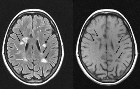
Accumulation of hypointense lesions (black holes) on T1 spin-echo MRI correlates with disease progression in multiple sclerosis.
Multiple sclerosis. We only use macrocyclic gadolinium-based contrast agents (as opposed to linear agents) due to its lower risk of associated gadolinium deposition in body tissues. tumefactive MS) are discussed separately. Geurts JJG, B L, Pouwels PJW, Castelijns JA, Polman CH, Barkhof F. 2005b. 16. One study at 1.5T using high-resolution 3D FLAIR showed only 5% of histologically confirmed cortical lesions, although this improved to 41% for mixed GM-WM (juxtacortical) and 71% for purely WM lesions (Geurts et al.
Patterns of enhancing lesion evolution in multiple sclerosis are uniform within patients. 2011; Lu et al. 2009. 2001;220(3):606-10. Diffusion tensor imaging of post mortem multiple sclerosis brain. 2015. Atrophy can be simplistically quantified in clinical settings by measuring ventricular width, corpus callosum area in cross-section, or intercaudate distance (Bakshi et al. In primary progressive MS (PPMS), cord abnormalities more than brain lesions are a hallmark of disease (Thorpe et al. 2016). One Hundred and Fifty Years Ago Charcot Reported Multiple Sclerosis as a New Neurological Disease. 2009; Ceccarelli et al. The .gov means its official.  Cortical lesions are nearly absent on conventional MRI sequences at lower field strengths (e.g., 1.5T) using standard resolution. Brain and spinal cord MRI lesions in primary An official website of the United States government. Tan I, van Schijndel R, Pouwels P et al.
 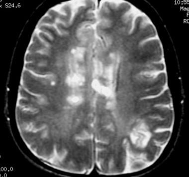 Imaging correlates of axonal swelling in chronic multiple sclerosis brains. 2012; Walsh et al. 2015. However, people with MS-like brain lesions that appear on an MRI scan have a 6080% chance of going on to develop another form of MS. With relapsing-remitting MS (RRMS), an MRI scan will show at least two separate areas of damage that have occurred at different points in time. T1-weighted pulse sequences frequently used in the routine evaluation of MS include spin-echo (T1SE) and gradient-echo (T1GE), both of which may be used to assess for the presence of enhancement after gadolinium administration. The introduction of magnetic resonance imaging (MRI) in the early 1980s revolutionized the diagnosis and treatment of multiple sclerosis (MS) by allowing unprecedented in vivo visualization of lesional activity and burden. Roberts, K. (2017). official website and that any information you provide is encrypted 2009. 2013).